Precision Targeting. Higher Detection. Localized Therapy.
Advanced Prostate Cancer Detection & Targeted Treatment
Detect. Target. Treat.
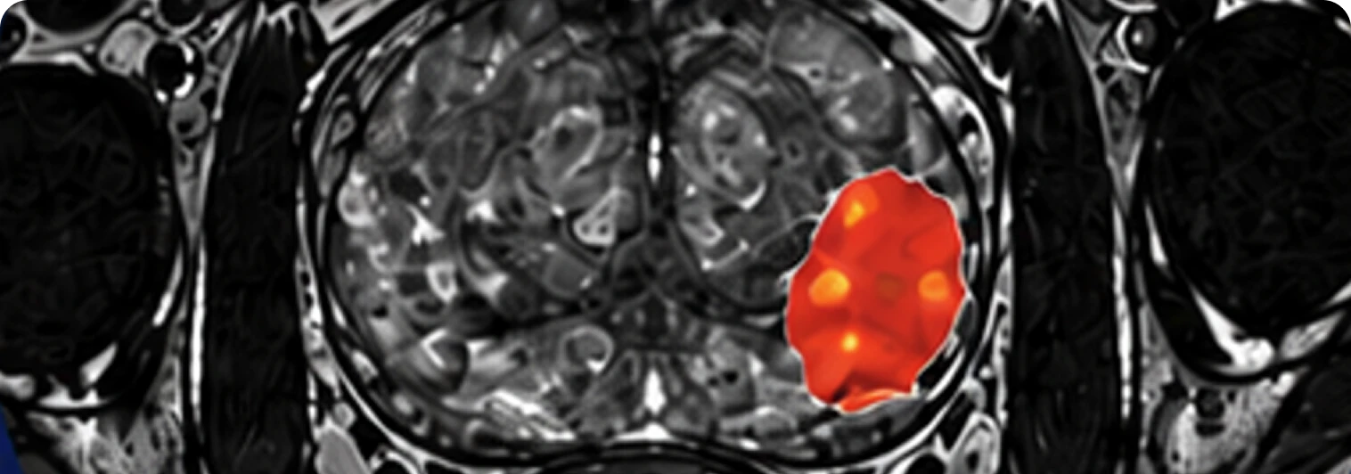
Detect
Advanced MRI identifies suspicious prostate lesions.
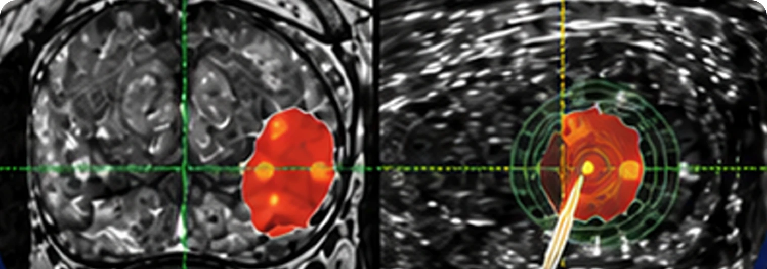
Target
MRI and real-time ultrasound are fused for precise targeting.
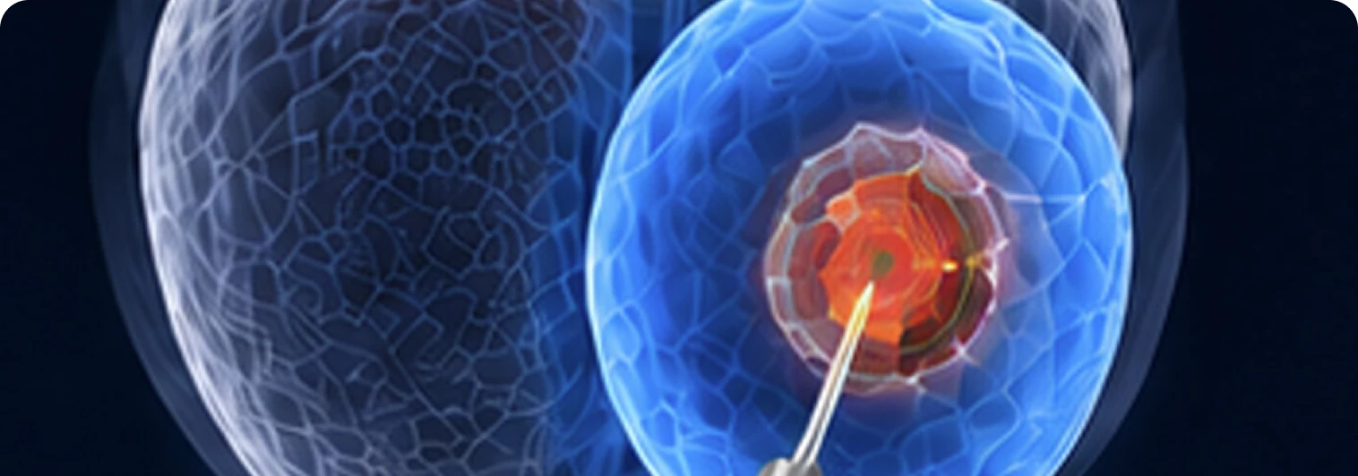
Treat
Precision-guided focal cryotherapy targets cancerous tissue while preserving healthy prostate tissue.
Clinical Procedural Support
Our Expertise

MRI Fusion Biopsy
MRI Fusion Biopsy fuses pre-acquired MRI images with real-time ultrasound, enabling physicians to precisely target and sample suspicious prostate lesions.
- MRI image integration and transfer
- Real-time MRI/ultrasound fusion
- Organ-based tracking technology
- 3D prostate mapping and documentation
- Follow-up and re-biopsy reference mapping
- In-office procedure

Focal Cryotherapy
Uses controlled freezing technology to precisely ablate cancerous tissue while preserving surrounding healthy structures.
- Complete cryotherapy platform and equipment
- Real-time ultrasound guidance throughout
- Treatment planning and contouring support
- Targeted tissue ablation
- Preservation of healthy tissue and function
- In-office procedure
Trusted Experience
25+
Years of Prostate Imaging Experience
20,000+
Procedures Supported
MRI Fusion Biopsy
Expertise
Focal Cryotherapy
Expertise
In-Office
Procedure Model
For Physicians
We integrate directly into your existing workflow, providing the equipment, technical support, and procedural expertise needed to perform advanced prostate procedures in your office.
For Patients
Our procedures are performed in the comfort of your physician's office using advanced image guidance designed to improve accuracy while minimizing disruption to your daily life.
OUR Technology
Advanced Technology. Precision Guidance.
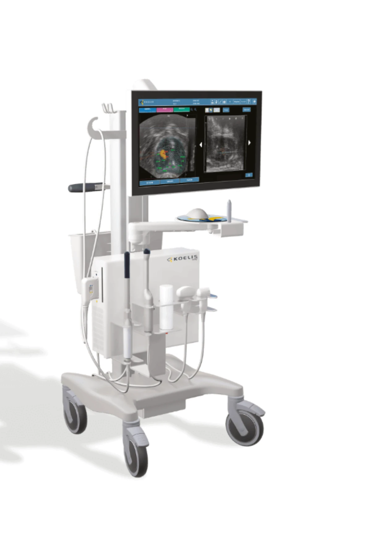
ADI utilizes the KOELIS Trinity® platform, an advanced MRI-ultrasound fusion system designed to enhance prostate cancer detection, biopsy targeting, treatment planning, and procedural accuracy. The mobile platform integrates seamlessly into the physician’s office, providing real-time imaging guidance and precise lesion localization throughout the patient journey.
3D MRI / Ultrasound Fusion
Combines MRI findings with real-time ultrasound imaging to accurately target suspicious prostate lesions.
Real-Time Imaging Guidance
Provides continuous visualization throughout biopsy and focal therapy procedures for greater confidence and precision.
Organ-Based Tracking Technology
Patented software adjusts for prostate movement during procedures, ensuring accurate targeting without extra tracking devices.
Office-Based Flexibility
Compact mobile design supports transperineal workflows in a convenient office-based setting.
3D Prostate Cartography
Creates a patient-specific three-dimensional map that can be used for biopsy documentation, active surveillance, treatment planning, and follow-up.
Proven Technology. Trusted Results.
The KOELIS Trinity® platform is clinically validated worldwide and used by leading urologists to improve accuracy and patient outcomes.
THE POWER OF AI-ENHANCED IMAGING
Higher detection rates.
Stronger clinical confidence.
OUR TECHNOLOGY
Al-Powered Imaging.
Smarter Decisions.
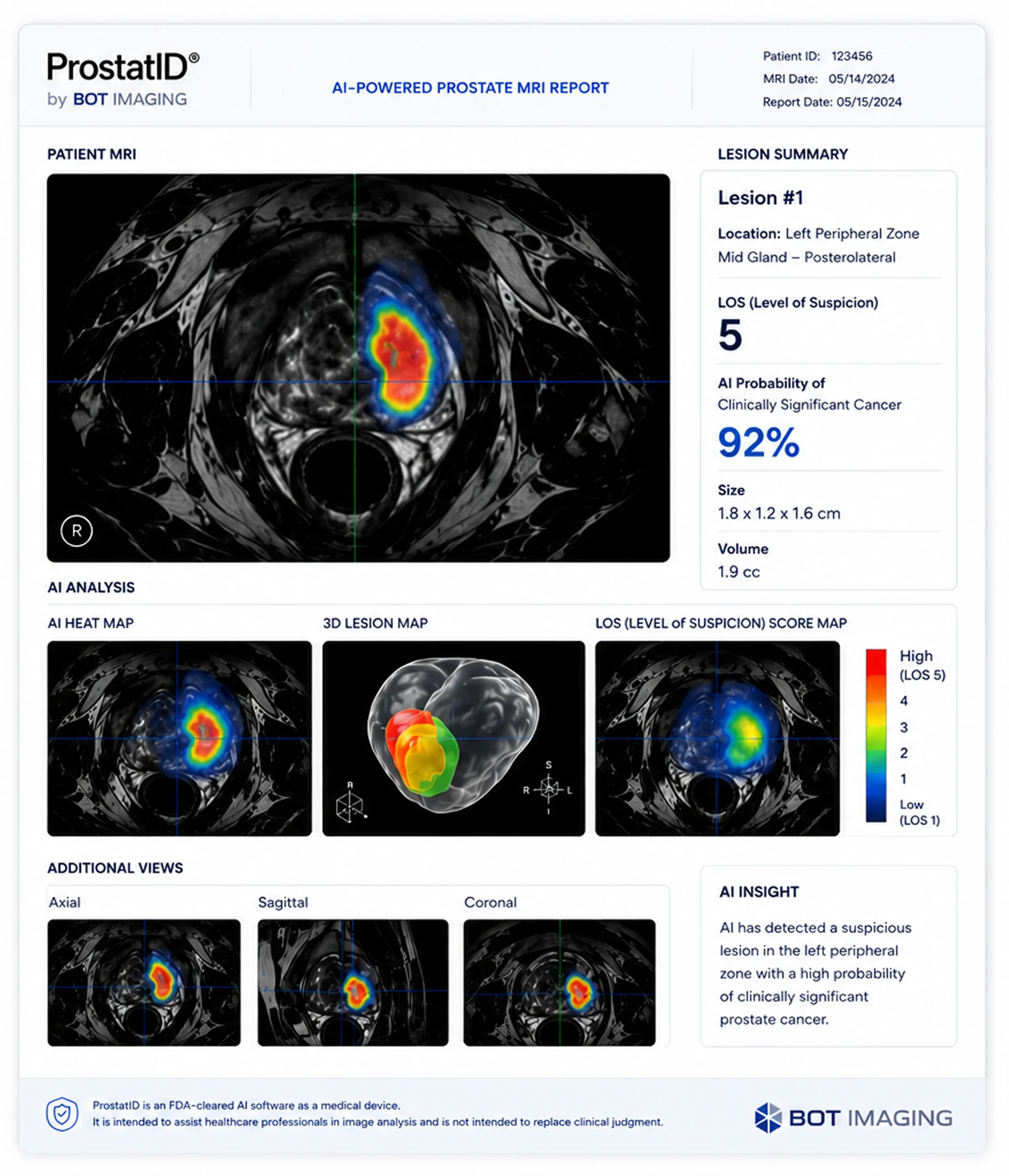
ADI utilizes BOT Imaging’s ProstatID®, an FDA-cleared artificial intelligence platform designed to assist physicians in the interpretation of prostate MRI.
ProstatID uses advanced deep-learning algorithms trained specifically on prostate imaging to identify and highlight areas suspicious for clinically significant cancer. It provides lesion localization, probability scoring, and 3D visualization to support clinical interpretation and targeted biopsy planning.
ProstatID is an advanced decision-support tool that complements physician expertise-not a replacement-helping improve confidence and consistency throughout the patient journey.
AI MRI Interpretation
Deep learning algorithms analyze MRI studies to identify suspicious lesions.
Lesion Detection & Localization
Automatically detects and localizes suspicious areas within the prostate.
Probability Scoring
Provides likelihood scores to support clinical decision-making.
3D Lesion Mapping
Creates 3D visualization for better understanding of lesion size and location.
Biopsy Planning Support
Works seamlessly with MRI fusion systems to improve targeted biopsy accuracy.
Smarter Imaging. Better Outcomes.
By combining advanced AI technology with expert clinical care, ADI helps physicians deliver more precise diagnoses and better outcomes for patients.
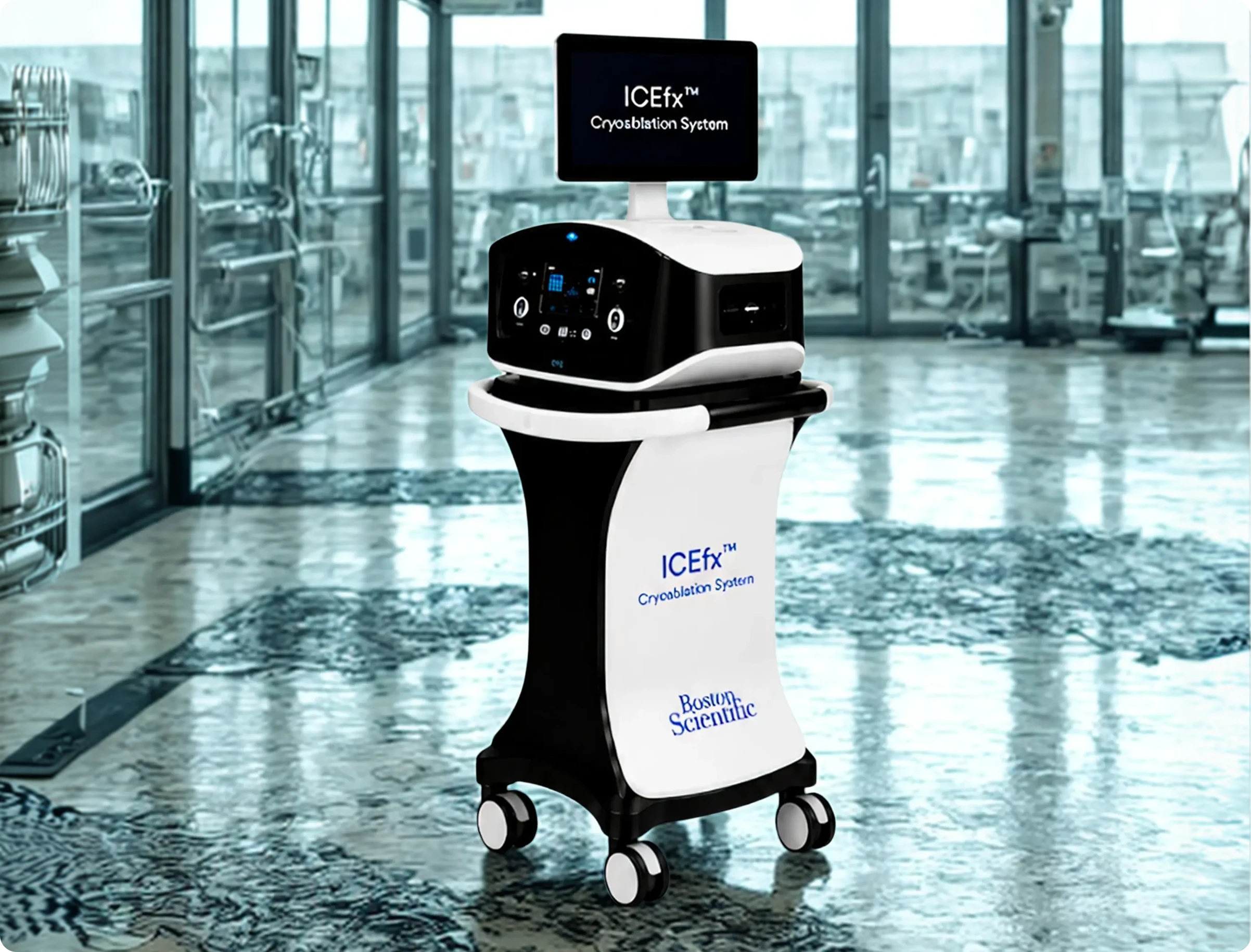
Image-Guided Cryotherapy
ADI offers the ICEv™ Cryoablation System to urology practices, providing advanced cryoablation technology that enables physicians to perform in-office cryotherapy procedures for prostate cancer in the office setting.
The ICEv™ Cryoablation System utilizes combined freezing technology and real-time imaging guidance to support precise, effective treatment.
Controlled Freezing
Precise crystallization technology
Real-Time Visualization
Continuous imaging throughout treatment
Targeted Treatment
Focused treatment zone with preservation of surrounding tissue
Real-Time Precision.
Targeted Results.
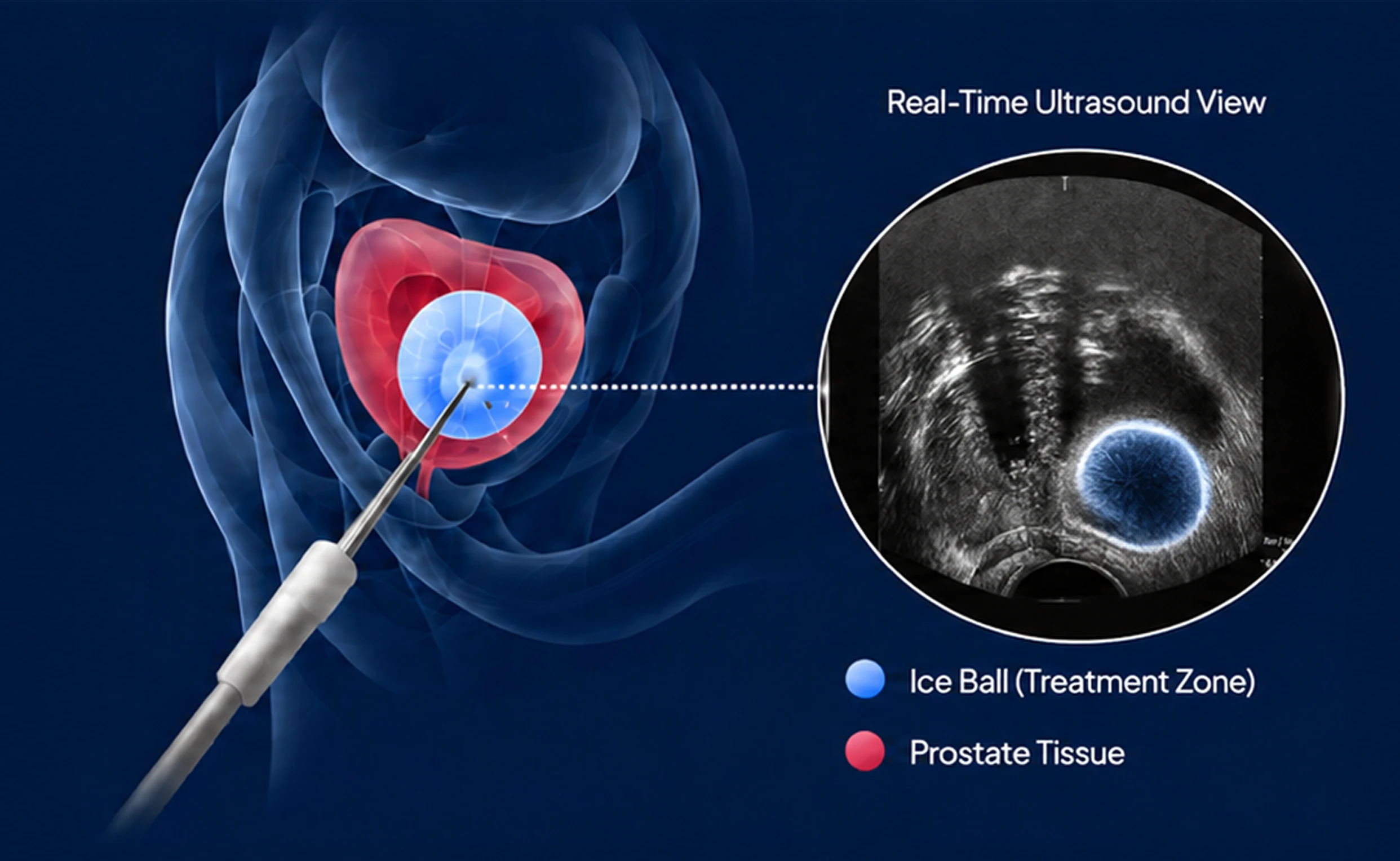
A thin cryo probe is carefully placed into the prostate under real-time ultrasound guidance. Argon gas cools the probe tip, forming an ice ball that conforms to the shape of the target.
The physician monitors the ice formation in real time to ensure precise coverage of the cancer while protecting critical structures.